
Canine Substitution for a Compromised Central Incisor
Ortho Insight, November 2025 · Dr John Sambevski, Specialist Orthodontist
An 11-year-old patient was referred for evaluation of a retained upper left deciduous canine and delayed eruption of the permanent successor. Imaging revealed transposition and impaction of the 23 into the root of the 21, with root resorption exceeding 50 per cent. This case discussion outlines the three treatment paths considered and the reasoning behind managing the case with canine substitution, avoiding the need for a prosthetic replacement.
Patient overview
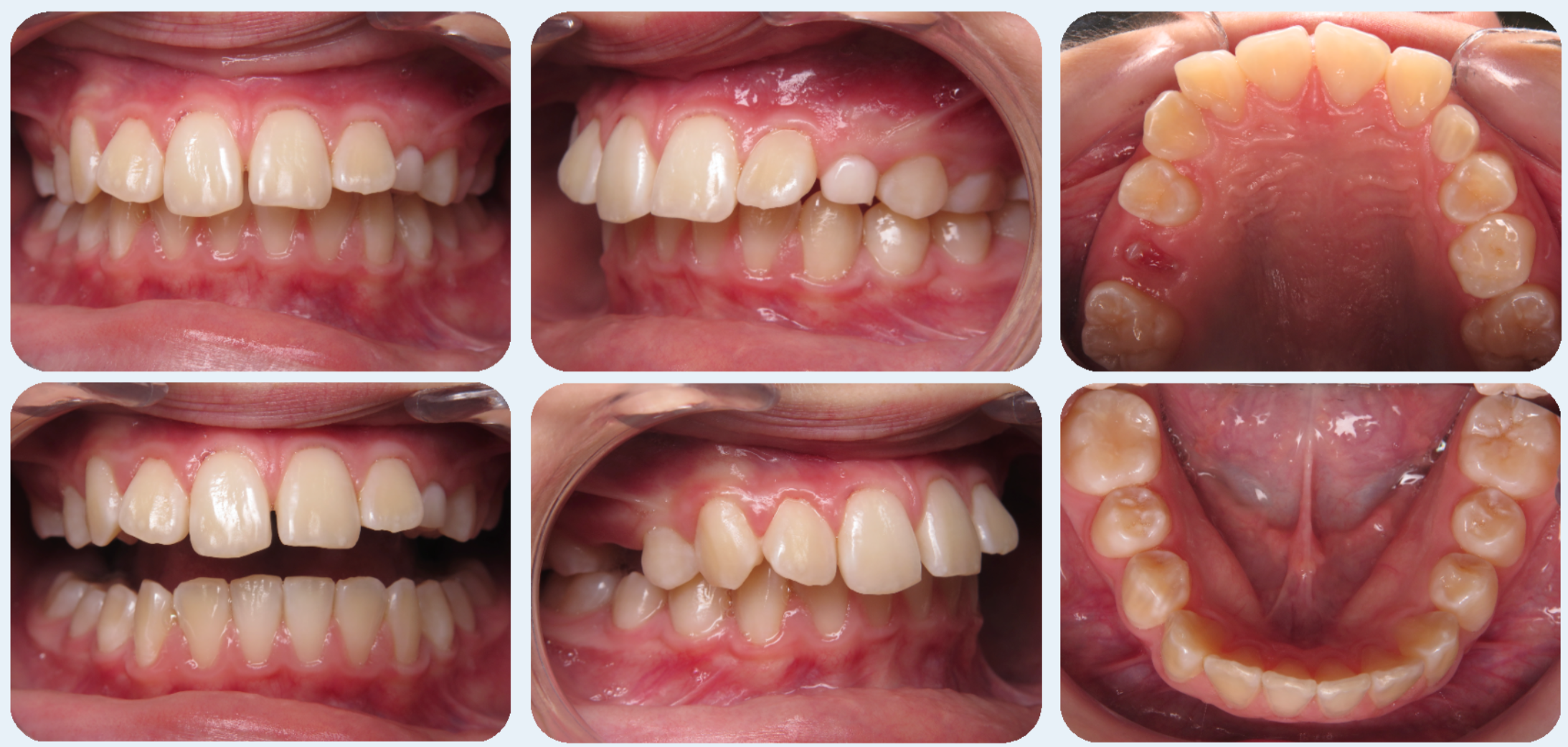
The patient presented with a Class II malocclusion and convex facial profile. Mild upper and lower crowding was noted, with increased overbite and overjet. No buccal canine bulge was palpable in the upper left quadrant, raising concern for palatal displacement of the 23. Early records highlighted an asymmetric eruption pattern, and further imaging was arranged to clarify the position of the 23.
Radiographic findings
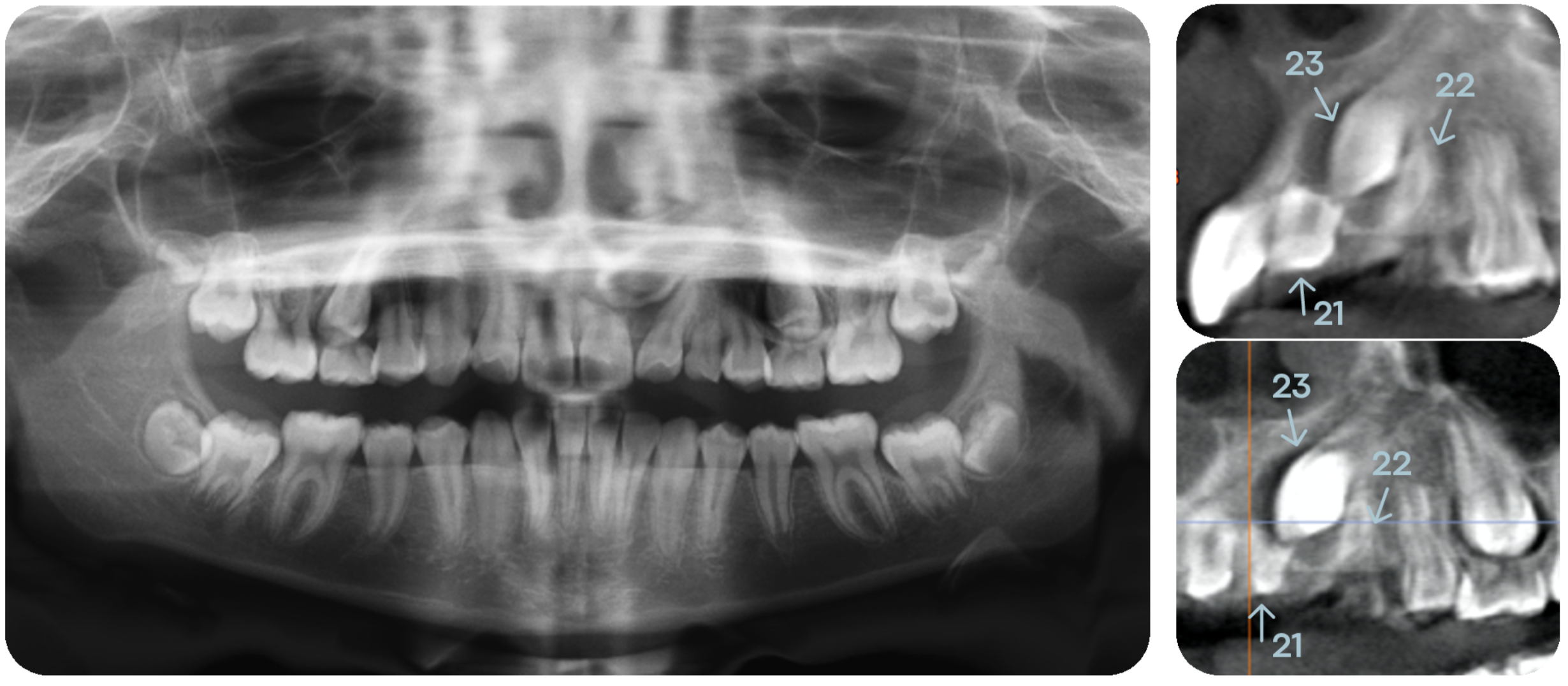
OPG and low-dose CBCT imaging revealed severe mesial displacement, transposition and impaction of the 23 into the root of the 21, resulting in more than 50 per cent root resorption of the 21 and distal displacement of the 22 root. Three-dimensional imaging was essential for assessing proximity, retrievability and the long-term prognosis of both the 21 and 23.
Treatment planning
Three treatment paths were discussed with the family:
1. Remove the 21, 23 and 63, with prosthetic replacement of the 21 by bridge or implant.
2. Retain the compromised 21 for as long as possible, extract the 23 and 63, and replace the 21 with an implant in the future.
3. Extract the 21 and 63, and allow the 23 to substitute for the 21, followed by reshaping to mimic a central incisor.
Given the poor prognosis of the 21, the family preferred the third, implant-free option.
Treatment progress
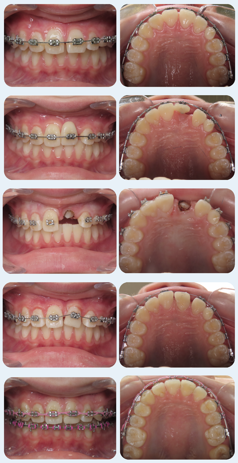
Treatment commenced on a non-extraction basis using upper fixed appliances. After initial alignment, the 21 was extracted by the referring dentist and the crown mounted to the archwire to maintain aesthetics during treatment. The impacted 23 was then surgically exposed and guided to erupt into the 21 position. A provisional composite build-up was added for aesthetics and space management. Lower appliances were placed to establish inter-arch mechanics, and the 63 was extracted to allow mesial movement of the posterior segment, which was managed without the need for TADs.
Outcome
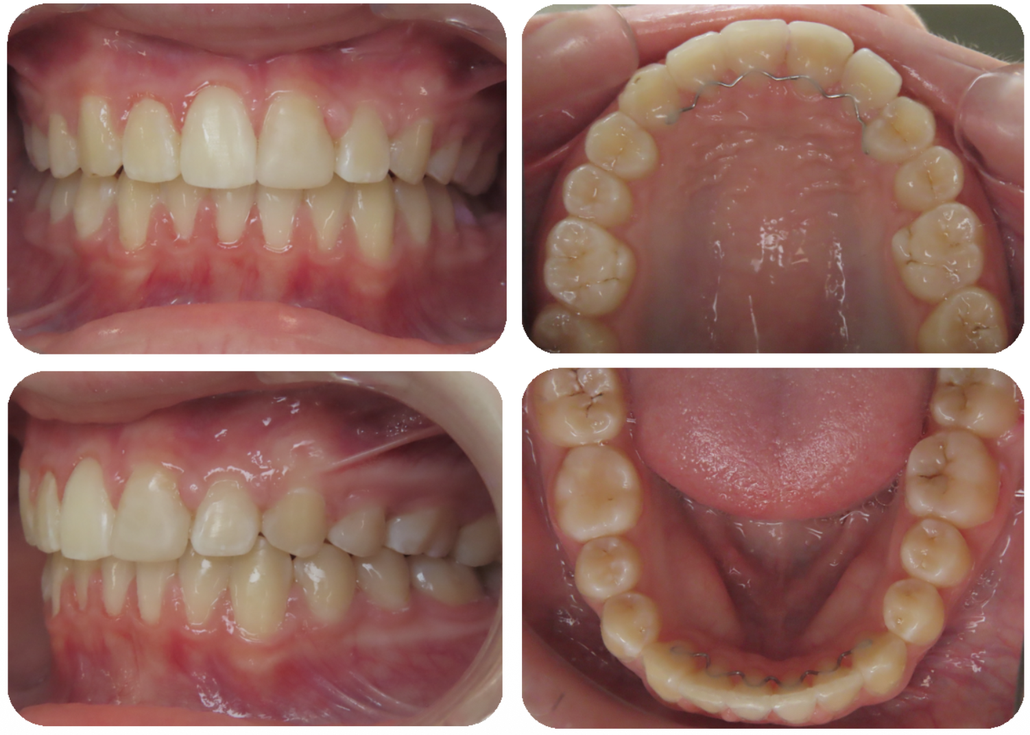
Treatment was completed after 40 months. The substituted 23 now functions in the position of a central incisor, following reshaping. The occlusion is stable, and minimal long-term maintenance is expected. The approach avoided a prosthetic replacement in a growing patient, which was central to the treatment planning discussion from the outset.
Technology notes
This case drew on a fully digital workflow: in-house low-dose CBCT for assessment of root morphology and impaction severity, 3D intraoral scanning for records and appliance design, and in-house 3D printing for model and retainer fabrication. In impaction cases, three-dimensional imaging materially changes the assessment of retrievability and prognosis, and it shaped the treatment options presented here.
Canine impaction: points for early detection
The prevalence of palatally displaced canines is estimated at 1 to 3 per cent in children, and ectopic eruption of maxillary canines affects about 2 per cent of the general population, occurring roughly twice as often in girls. Evidence of possible impaction can be apparent from age 9, peaking around ages 11 to 13. Common causes include insufficient arch space, delayed or early loss of primary teeth, and abnormal development of adjacent teeth. Early detection and referral matter because impaction and abnormal eruption can lead to crowding, root resorption of adjacent teeth, and misalignment. An absent buccal canine bulge by age 9 to 10 is a practical chairside prompt for further investigation.
Canine transposition
Maxillary canine transposition has a reported prevalence of 0.14 to 0.51 per cent, appearing far more frequently in the maxilla than the mandible. In orthodontic patient populations the rate is around 0.5 per cent. The most common transpositions are between the maxillary canine and first premolar, accounting for about 71 per cent of cases, and between the maxillary canine and lateral incisor, about 20 per cent.
We thank our referring colleagues for their trust and collaboration in managing complex eruption cases. All records and photographs are used with the patient's permission.
This case was originally published as Ortho Insight, November 2025, printed and posted to our referring practices. [Download the print edition (PDF)]